For Patients: Dental Implant Failure Checklist, 2–7% Need Replacement
Yes, dental implants can fail. Roughly 2 to 7 percent do, depending on the study and how long it’s been tracked. If yours feels loose, hurts, or looks different than it did last month, stop chewing on that side, call your dentist today, and pull together your recent x-rays and surgical notes. Most causes are identifiable, and a staged treatment plan exists even after failure.
TL;DR:
- Early implant failures, mostly within the first year, are linked to poor osseointegration caused by low bone density, infection, or surgical trauma.
- Most failures result from peri-implantitis, a gum infection leading to bone loss, especially in patients with a history of periodontal disease.
- Late failures often occur due to mechanical overload, bruxism, or poor prosthetic design, rather than infection.
- Risk factors like smoking, uncontrolled diabetes, certain medications, and systemic health conditions significantly influence failure odds.
- Regular imaging, symptom monitoring, and personalized risk assessment help improve diagnosis, prevention, and treatment success.
Table of Contents
- What Does Dental Implant Failure Actually Mean?
- How Common Is Implant Failure, Really?
- Why Peri-Implantitis Drives Most Biological Failures
- When the Problem Is Mechanical, Not Infectious
- Systemic and Medication Risk Factors Worth Discussing Before Surgery
- Signs Your Implant May Be Failing, Ranked by Urgency
- What Happens at the Dental Visit: Exam and Imaging
- The Treatment Pathway: From Conservative Care to Removal
- Life After Implant Failure: Replacement and Realistic Timelines
- How Implant Veneer Guide’s Tools Fit Into This Process
- A Compact Checklist Before Your Next Appointment
- The Emotional Weight of a Failing Implant
- Why Your Overall Health Changes the Odds
- The Perspective Missing From Most Implant Guides
- Plan Your Next Step With the Right Tools
- Sources
What Does Dental Implant Failure Actually Mean?
“Dental implant failure” isn’t one condition. It’s a category that covers two very different problems, and mixing them up is why so many patients panic unnecessarily or, worse, ignore something urgent.
The first type is a failure of osseointegration, meaning the titanium post never properly fused with the jawbone, or it did fuse and later lost that bond. The second is prosthetic failure, where the implant itself is solidly anchored but the crown, abutment, or screw on top breaks, loosens, or wears out. A cracked crown is an annoyance you fix in one visit. A failed integration is a structural problem that may require removing the implant entirely.
Timing splits failures into two buckets clinicians treat differently:
- Early failure happens before or during the loading phase, typically within the first few months, and usually points to a healing problem: infection, poor bone quality, or the implant being disturbed before it fused.
- Late failure occurs after the implant has been chewing food successfully for months or years, and it’s almost always a maintenance, inflammation, or mechanical overload issue.
Why does this distinction matter so much? An early failure often means the site needs to heal and can be reattempted later. A late failure, especially one caused by advancing bone loss around the implant, is more likely to threaten neighboring teeth and requires faster intervention. Your dentist’s first question when you report a problem is usually “how long has this implant been in function?” because the answer reshapes the entire treatment conversation.
How Common Is Implant Failure, Really?
Implant failure is uncommon, but it isn’t rare enough to dismiss. A large retrospective cohort covering 158,824 implants found an overall failure rate of 2.21%, and a broader scoping review of 388 studies put early failures somewhere between 0.5% and 5.2%, with late failures ranging from 0.5% to 7.8%.
The numbers that matter most: in the 158,824-implant cohort, 1.59% of all implants failed within the first year alone, and failure rates ran higher in certain maxillary (upper jaw) sites, closer to 3%. That’s not a coincidence. Upper jaw bone tends to be softer and less dense than lower jaw bone, which gives implants less to grip during the critical healing window.
Two patterns hold up across most of the literature. First, failures cluster early. The first twelve months carry the highest single-year risk, since that’s when osseointegration either succeeds or doesn’t. Second, there’s often a smaller secondary wave around the three-year mark, which tends to reflect peri-implant disease that’s had time to develop rather than a healing problem.
Location and prosthesis type both shift the odds. Implants supporting full-arch bridges or All-on-4 restorations distribute load differently than single-tooth implants, and multi-unit prosthetics can mask an individual implant’s struggles until the whole restoration starts to feel off. If you’ve had implants placed in the upper back jaw, where bone is typically thinner, it’s worth asking your dentist directly what site-specific risk they factored into your treatment plan.
Why Peri-Implantitis Drives Most Biological Failures
Peri-implantitis is the single most cited biological cause of implant failure in the clinical literature, showing up in roughly 150 of the 388 studies reviewed in one major scoping review. It’s essentially gum disease around an implant instead of a natural tooth, and it behaves aggressively because implants lack the natural fiber attachment that slows infection around real tooth roots.
The clinical picture is fairly consistent:
- Redness, swelling, or puffiness in the gum tissue surrounding the implant
- Bleeding when the site is probed or even brushed
- Pus or discharge, sometimes only visible when the gum is pressed
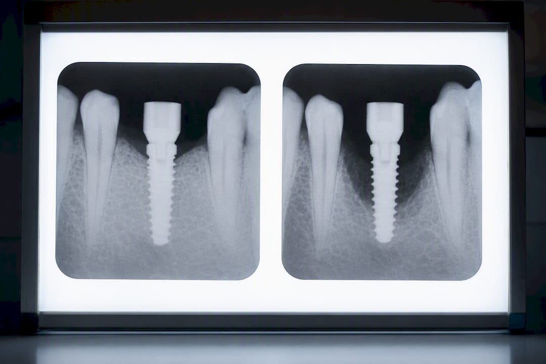
- Progressive bone loss visible on x-rays, which is what separates peri-implantitis from milder peri-implant mucositis (inflammation without bone loss yet)
Patients with a documented history of periodontitis face meaningfully higher risk. That history signals the mouth’s bacterial environment and immune response already struggled to control gum disease around natural teeth, and implants don’t get a pass from that same biology.
Early osseointegration failure has its own set of biological triggers, distinct from peri-implantitis. A retrospective analysis of implant removals found that lack of osseointegration accounted for 36.4% of failures and absence of primary stability for another 22.4%, meaning more than half of removals traced back to the implant simply never gripping the bone properly in the first place. Contributing factors include low-grade infection introduced during surgery, insufficient bone density at the site, and surgical trauma from overheating the bone during drilling.
There’s also an emerging, still-developing area of research into implant material itself. Some studies suggest that titanium particle release from the implant surface may provoke an inflammatory response in susceptible patients, though this hasn’t been established as a major clinical driver and shouldn’t be a primary worry for most people.
Pro Tip: Ask your dentist to show you your baseline post-op x-ray and compare it side by side with your most recent one. Bone loss is easiest to catch as a trend across images, not from a single snapshot.
When the Problem Is Mechanical, Not Infectious
Not every failing implant is fighting an infection. A significant share of late failures come down to physics: too much force, applied in the wrong direction, for too long.
Occlusal overload happens when an implant absorbs more chewing force than it was designed to handle, often because the bite wasn’t balanced correctly at the time of restoration, or because the patient’s bite has shifted since. Bruxism, the habit of grinding or clenching teeth (often overnight and unknowingly), is one of the most common accelerants here. Natural teeth have a ligament that cushions bite force. Implants don’t, so that force transmits directly into the bone, which over years can trigger microfractures or loosen the implant-bone interface.
Prosthetic design contributes its own set of problems, and this is one dentists underdiscuss:
- Crowns or bridges with overhanging edges that trap plaque and are nearly impossible to floss around
- Restorations placed at an angle that makes daily cleaning around the gumline unrealistic
- Screw-retained crowns with loosening abutment screws, which mimic the pain and mobility of a genuinely failing implant
That last point matters more than most patients realize. A loose abutment screw can feel exactly like a loose implant. The fix might be a five-minute screw retightening rather than surgery, but you won’t know which one you’re dealing with until a dentist examines it. If a crown suddenly feels like it’s rocking, resist the urge to assume the worst before you’ve had it checked.
Systemic and Medication Risk Factors Worth Discussing Before Surgery
Some of the strongest predictors of implant failure have nothing to do with the mouth directly. They’re systemic, and many are modifiable if you catch them early enough in the planning process.
Smoking is the highest-impact behavioral risk factor identified across the implant literature. Nicotine restricts blood flow to healing tissue and slows bone formation, which directly undermines osseointegration during the critical early months. Uncontrolled diabetes runs a close second. Blood glucose that stays elevated impairs wound healing and increases infection susceptibility, both of which stack the odds against a new implant.
Several medication classes have documented associations with altered bone metabolism, though the strength of evidence varies:
- Bisphosphonates, used for osteoporosis, are linked to impaired bone remodeling and, in rare cases, medication-related osteonecrosis of the jaw
- SSRIs (a common antidepressant class) have been associated in some studies with reduced bone density, though causation isn’t firmly settled
- PPIs (proton pump inhibitors for acid reflux) may interfere with calcium absorption over long-term use, with evidence still evolving
Clinical consensus recommends more intensive maintenance, often 3 to 4 professional cleanings per year, for patients with a periodontitis history, since that background risk doesn’t disappear once an implant is placed.
None of this means these medications rule out implants. It means your dentist needs your full medication list before surgery, not just your dental history. If you’re managing diabetes, ask specifically what HbA1c target your surgical team wants to see before scheduling, since most protocols aim for a stable range rather than a single number. If you’re on bisphosphonate therapy or have osteoporosis, that conversation should happen well before the surgical date, not the week of.
Signs Your Implant May Be Failing, Ranked by Urgency
Some warning signs are impossible to miss. Others are subtle enough that patients dismiss them for months. Here’s how to read your own symptoms:
- Visible mobility. If you can feel the implant crown move when you press on it with your tongue or a finger, that’s the clearest red flag there is. Call your dentist the same day.
- Pain that’s new or worsening. Implants shouldn’t ache once healed. New pain, especially throbbing or pain that wakes you at night, suggests active infection or inflammation.
- Swelling, pus, or a bad taste near the site. Any discharge is a sign of infection and needs evaluation within days, not weeks.
- Bleeding gums around the implant, particularly when brushing or flossing, even without pain.
- A hollow or dull sound when tapped. Dentists sometimes tap implants during checkups; a hollow percussion note can indicate the implant has lost its solid bone connection even before mobility is obvious.
- A change in your bite. If your teeth suddenly meet differently, or the implant crown feels “tall” compared to neighboring teeth, that can signal bone loss shifting the implant’s position.
- Gum recession exposing metal. If you can see the gray or silver implant collar where you couldn’t before, the supporting tissue is receding.
Numbers 1 through 3 warrant a same-week appointment at minimum. Numbers 4 through 7 are worth flagging at your next visit but should still prompt a call if they persist more than a couple of weeks. For a deeper breakdown of what these signs of implant failure typically indicate, it’s worth reading before your appointment so you can describe symptoms precisely.
What Happens at the Dental Visit: Exam and Imaging
Diagnosing a failing implant follows a fairly standard sequence, and knowing it ahead of time makes the appointment less stressful.
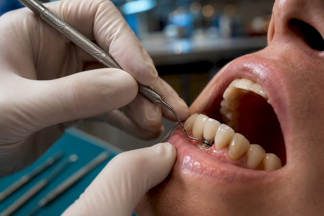
Your dentist starts with a periodontal probe, gently measuring the depth of the pocket around the implant in several spots. Bleeding on probing is a key signal; a healthy implant site typically doesn’t bleed when probed gently. Suppuration, meaning visible pus when pressure is applied, confirms active infection rather than simple irritation. Mobility testing follows, usually done by hand or with an instrument, since any detectable movement in a mature implant is abnormal.
Imaging comes next:
- Periapical x-rays are the standard first step, giving a two-dimensional view of bone levels around the implant compared against your baseline post-surgical image.
- CBCT (cone beam CT) scans provide a three-dimensional view and are typically reserved for cases where bone loss looks complex, asymmetrical, or where surgical planning for pretreatment is already underway.
If probing depths exceed a certain threshold combined with confirmed bone loss on imaging, or if the case involves complex anatomy near the sinus or nerve canal, your general dentist will usually refer you to a periodontist or oral surgeon. That referral isn’t a sign of a worst-case scenario. It’s standard practice once a case moves from monitoring into active treatment decisions, and specialists have more tools and more experience with the surgical side of implant salvage.
The Treatment Pathway: From Conservative Care to Removal
Treatment for a failing implant follows a staged approach, moving from the least invasive option to the most invasive only when necessary.
Non-surgical management comes first whenever the case allows it. This usually means mechanical debridement to clear bacterial buildup around the implant surface, combined with local antiseptic rinses (chlorhexidine is common) or localized antibiotic delivery directly into the pocket. This tier works best for early-stage inflammation without significant bone loss.
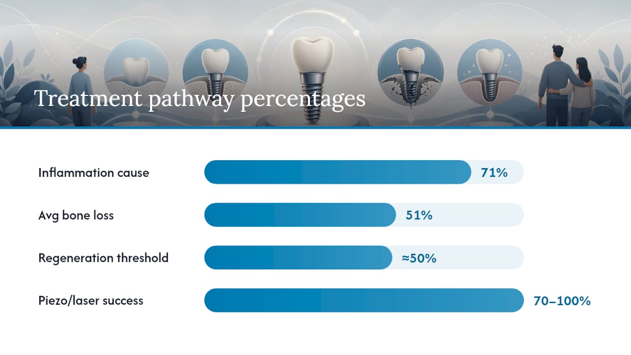
Surgical therapy becomes necessary once bone loss is confirmed. Open flap debridement lets the surgeon directly access and clean the implant surface, sometimes paired with regenerative techniques like bone grafting material to encourage new bone growth around the exposed implant threads. Success here depends heavily on how much bone has already been lost. Once a case crosses roughly 50% crestal bone loss, outcomes for regenerative therapy drop sharply, which is often the threshold clinicians use when deciding retention isn’t realistic anymore.
Explantation is the last step, reserved for implants that can’t be salvaged. A retrospective cross-sectional study of 738 implant removals found peri-implant inflammation was responsible for 71% of cases, with average bone loss at the time of removal around 51%.
Removal technique matters for what comes next:
- Trephine burs remove a cylinder of bone along with the implant, which is effective but almost always requires bone grafting afterward.
- Reverse-torque devices unscrew the implant using counter-rotation, which can preserve more surrounding bone when the implant hasn’t fully fused.
- Piezoelectric and laser-assisted techniques are gentler on soft tissue and bone, with reported success rates ranging from 70% to 100% depending on case complexity, though they require specialized equipment not every practice has.
Pro Tip: Ask which explantation method your surgeon plans to use and why, before the day of the procedure. The technique directly affects how much grafting you’ll need afterward and how long you’ll wait before a new implant is even possible.
Life After Implant Failure: Replacement and Realistic Timelines
Losing an implant isn’t the end of the road, but it does reset the clock, and knowing what to expect helps you plan rather than panic.
Re-implantation at the same site is often possible, but timing depends entirely on how much bone was lost. If minimal grafting was needed, some surgeons will consider a new implant in as little as 3 to 4 months. If significant grafting or a sinus lift was required, that window commonly stretches to 6 to 9 months or longer, since new bone needs time to mature and integrate before it can support another implant.
Your alternatives while you wait, or if re-implantation isn’t advisable, generally fall into three categories:
- Fixed bridges anchored to adjacent natural teeth or implants, which restore function without needing bone at the failed site.
- Removable overdentures, a lower-cost option that rests on the gum and remaining structures, though patients often find them less stable than fixed solutions.
- All-on-4 style full-arch restorations, which can sometimes bypass a problem site entirely by anchoring the prosthesis to implants placed in stronger bone elsewhere in the jaw.
Bone grafting and sinus lifts, when needed, add real time to any of these paths. A sinus lift alone commonly needs 4 to 6 months of healing before it can support an implant reliably. This is exactly the kind of timeline confusion that recovery planning resources are built to clarify, since patients frequently underestimate how long staged treatment actually takes.
How Implant Veneer Guide’s Tools Fit Into This Process
Clinical judgment always comes first, but a few structured tools can sharpen the conversation you have with your dentist.
The Dental Implant Candidacy Checker walks through the systemic and behavioral risk factors covered above, smoking status, glycemic control, bone density history, so you arrive at a consultation already knowing which questions matter for your case. It’s a screening aid, not a diagnosis; only a clinical exam and imaging can confirm candidacy.
Once you’re weighing replacement options, the Cost Estimator and Recovery Timelines help translate clinical terms like “grafting” and “sinus lift” into realistic budget ranges and healing windows, so you’re planning with numbers instead of guesses.
Every guide on the platform is built from peer-reviewed sources and cross-checked against current clinical literature, but none of it replaces an in-person exam. Treat these as preparation tools for better conversations, not substitutes for the conversation itself.
A Compact Checklist Before Your Next Appointment
Before you walk into your dentist’s office, get organized. It changes how efficiently the visit goes.
- Photograph the site daily for a few days if symptoms are mild, so you have a visual timeline
- Write down when symptoms started and whether they’re constant or triggered by chewing
- List every medication and supplement you’re currently taking, including over-the-counter ones
- Bring or request your original surgical records and the most recent x-ray on file
At the appointment, ask these six questions directly: What’s the likely cause? Is this implant salvageable? What imaging do you need to confirm that? What’s the realistic timeline if treatment starts today? What replacement options exist if it fails? And what maintenance schedule prevents this from happening to my other implants? A risks and complications overview can help you understand the answers you get back.
The Emotional Weight of a Failing Implant
Implant failure isn’t just a clinical event. For a lot of patients, it’s a genuine blow, especially after months of surgery, healing, and cost that felt like they’d already paid the hard price once.
It’s common to feel frustration, even anger, when a problem surfaces after you’d mentally filed the procedure as “done.” Some patients describe a kind of grief specific to dental work: the sense that their body rejected an investment they made in themselves. Others spiral into anxiety about their remaining implants, checking them obsessively or avoiding certain foods long after a dentist has cleared them to eat normally.
None of that reaction is irrational. Dental work is expensive, invasive, and tied up with appearance and confidence in ways that other medical procedures often aren’t. Reading firsthand accounts, including the more difficult ones documented in pieces like what patients need to know about implant complications, can validate that frustration without pretending every case ends badly.
The most useful coping strategy isn’t forced positivity. It’s information. Understanding why an implant failed, whether it was preventable, and what the actual odds are for a successful replacement tends to lower anxiety more effectively than reassurance alone. If the emotional impact is significant, particularly around eating, socializing, or self-image, it’s worth mentioning to your dentist directly. Many practices coordinate with brief counseling support for patients navigating a difficult treatment course, and there’s no reason to manage that weight alone.
Why Your Overall Health Changes the Odds
Implant failure risk doesn’t exist in isolation from the rest of your health. Systemic conditions shift both the odds of failure and, just as importantly, the odds of a successful second attempt.
Diabetes is the clearest example. Well-controlled diabetes, with HbA1c in a stable target range, carries a risk profile not far from that of a non-diabetic patient. Poorly controlled diabetes changes that picture substantially, since chronic high blood sugar impairs the microvascular healing implants depend on. This is why most surgical protocols now require a recent HbA1c reading before scheduling, not as a formality but because it genuinely predicts healing capacity.
Osteoporosis and other bone-density conditions matter for a different reason: they can affect how well bone integrates around the implant in the first place, particularly in the jaw regions already prone to thinner bone. Autoimmune conditions and certain cancer treatments that suppress the immune system also slow healing and raise infection risk during the vulnerable early months.
None of these conditions are automatic disqualifiers. What they change is the conversation. A patient with well-managed diabetes or stable osteoporosis under a physician’s care can often proceed with implants using extra precautions: staged healing timelines, closer follow-up intervals, and sometimes a preference for grafting techniques with a stronger track record in compromised bone. The mistake is treating implants as a purely dental decision when, for a meaningful share of patients, the medical history is doing as much of the work as the surgical technique.
The Perspective Missing From Most Implant Guides
Most implant content treats failure like a footnote, mentioned once, then buried under glowing success-rate statistics. That’s backward. Patients don’t need to be scared away from implants; they need to understand that a 2 to 7 percent failure range is real enough to plan for, not a rounding error.
The conventional advice, “brush well and see your dentist twice a year,” undersells what actually predicts outcomes. The data points somewhere more specific: glycemic control, smoking status, and whether your restoration is designed for you to actually clean it daily matter more than generic hygiene advice ever will. A beautifully placed implant under a poorly designed crown will fail on a predictable timeline, and that’s a design problem, not a brushing problem.
If there’s one priority worth acting on, it’s this: get your baseline x-ray and keep it. Every future comparison depends on having that reference point, and too many patients don’t request one until something already feels wrong. Ask for it before you leave your surgical follow-up, not after your implant starts hurting.
— Georgina
Plan Your Next Step With the Right Tools
Reading about causes and treatment pathways only gets you so far. What actually helps is putting your own risk factors, health history, and budget into a structured framework before you sit down with a dentist.
The Dental Implant Candidacy Checker turns everything covered here, smoking status, glycemic control, bone density history, into a personalized screening summary you can bring straight into a consultation. It won’t diagnose anything; only an exam and imaging can do that. But it does mean you walk in already knowing which risk factors apply to you, instead of discovering them mid-appointment.
If you’re weighing what a replacement or full-arch alternative might cost, the Cost Estimator gives realistic US ranges by procedure type, and the Patient Journey tool maps out what each stage, from consultation through long-term follow-up, actually involves. Both are planning aids built from published clinical data, not a substitute for your dentist’s evaluation. Start with the Candidacy Checker, then bring your results to your next appointment.
Sources
The claims in this guide draw directly from peer-reviewed sources you can read in full:
- Large cohort analysis of implant survival (Maccabi Dent database)
- Timing and extent of peri-implant bone loss for dental implant removal